Original Article
Probability of Hereditary Effects Due to Dose Radiation on X-Ray Radiography Examination
I Gusti Agung Ayu Ratnawati, Ida Bagus Made Suryatika, Gusti Ngurah Sutapa, Anak Agung Ngurah Gunawan*
Department of Mathematics and Natural Sciences, Udayana University, Badung 80361, Bali, Indonesia
*Corresponding Author’s Email: a.a.ngurahgunawan@unud.ac.id
Abstract
Introduction: This study estimates the risk of hereditary diseases related to X-ray radiation in conventional radiography, conducted at the Bali Academy of Radiodiagnostic and Radiotherapy Engineering using a Raysafe Multimeter. The focus is on stochastic radiation effects, including hereditary and cancerous impacts. These effects, which occur without a specific dose threshold, are linked to mutations in somatic and germinal cells that contribute to cancer and genetic disorders. The goal was to assess the hereditary risk by measuring radiation doses through the Entrance Surface Dose (ESD). Methods: Key variables measured during the radiographic procedure included output voltage (kV), exposure time (s), current output (mA), and radiation dose exposure (mGy). These measurements were used to calculate the ESD, which helped estimate hereditary risks for critical organs. Data collected with the Raysafe Multimeter was analyzed to establish the relationship between radiation dose and hereditary risk. Results: The study found that the lowest ESD value was 0.3737 mGy with 40 kV voltage, and the highest was 0.7328 mGy at 80 kV. It also showed that the first generation (Generation I) had a higher probability of hereditary effects than the second generation (Generation II). This was attributed to the cells' ability to repair radiation-induced damage over time, with the second generation benefiting from more opportunities for repair. Conclusion: The risk of hereditary diseases is influenced by the ESD and the cell’s repair ability. The second generation faces lower hereditary risk due to natural repair processes, underscoring the need for careful radiation exposure management in radiography.Keywords: Absorbed Dose; Critical Organs; ESD; Hereditary Risk; Radiation Exposure
Introduction
Conventional radiography is an examination that uses a conventional X-ray that is installed permanently or mobile in a room that is used for routine general examinations. The radiation source used for this examination is X-rays (Reis et al., 2023). The advantages of X-rays such as body imaging technology, without surgery, and providing a detailed anatomical picture are the benefits of X-rays. Diagnostic radiology is the most common practice of utilizing X-ray radiation, and there has been a substantial increase in the number of examinations (Rayan et al., 2025). The introduction should clearly summarize the purpose and rationale of the study without extensively reviewing the subject or presenting data or conclusions (Utami et al., 2024).
Based on the principle of radiographic formation, it is possible to receive radiation exposure to the object that is passed, in this case, the human body. Factors that affect the quality of radiography are exposure factors consisting of tube voltage (kV), tube current (mA), and exposure time(s). So that exposure factors can also affect the occurrence of radiation effects (Imaoka et al., 2023). The stochastic effect is a radiation effect that has a chance, along with exposure to radiation that hits biological organs. This stochastic effect does not have a specific dose threshold (Stewart et al., 2012). Examples of stochastic effects are cancer, leukemia, and others (Mothersill et al., 2024). Non-stochastic effects or deterministic effects are also called definite effects because they do not follow the function of probability. The deterministic effect occurs when a certain dose threshold has been exceeded. However, this effect occurs immediately once the threshold dose is exceeded. As an example of deterministic effects, namely sterilization (sterilization), burns (erythema), cataracts, and fetal death (teratogenic) (Romodin et al., 2025).
Hereditary effects are mutations that occur naturally or spontaneously in the somatic and germinal cells, respectively, contributing to the induction of cancer and inherited genetic diseases. The study of the genetic effects caused by radiation is much more difficult than the study of cancer (Choudhuri et al., 2021). Among other things, due to the lack of information about damage to human genetic material due to radiation, mutations, which are induced by radiation, are recessive, so there is a possibility that they cannot be detected for several generations later (Rajeev et al., 2026). It takes several generations for the effects to occur. Long generation periods in humans, lack of dose data, few known populations are exposed to significant amounts of radiation, and the genetic effects induced by ionizing radiation are indistinguishable from other causes (Tandon & Srivastava, 2025).
The patient's dose assessment on a general radiographic examination can be expressed in the Entrance Surface Dose (ESD) (Alomairy et al., 2023). Indirect ESD measurement is considered to be the most technically feasible method (Kanzaki et al., 2023). The indirect ESD measurement method can be done with an approach based on the tube output of each tool (Liu et al., 2024). This study aims to estimate the dose profile of patients undergoing general radiography examination using the tube output measurement method, then the probability of heredity effects from generation to generation can be determined (Suliman, 2020).
Methodology
Study Setting and Equipment
This research was carried out at the Academy of Radiodiagnostic and Radiotherapy Engineering (ATRO) Bali using an X-ray aircraft brand Medical Instrument System (MIS) type/model MXHF-1300R. Method of measuring the tube output of X-ray aircraft of patients undergoing general radiography examination with a Raysafe Multimeter (Andriati et al., 2024).
Radiographic Examination Parameters
The irradiation condition is carried out at tube voltages of 50, 60, 70, 80, and 90 kV with an irradiation time of 0.1 seconds and a tube current of 100 mA. The series of irradiation implementation can be shown in Figure 1 below,
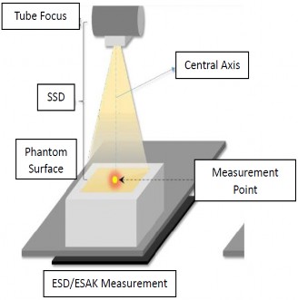
Figure 1: Entrance Surface Dose (ESD) Measurement (da Silveira et al., 2026)
Radiation Measurement Process
ESD calculations were carried out at the ATRO Bali diagnostic laboratory using a Raysafe Multimeter. From the Raysafe Multimeter, the output value of the X-ray plane is generated, with the unit mGy converted to μGy. After obtaining the output value of the aircraft with μGy units, then the output value of the aircraft is divided by the mAs used in the research. The output value/mAs, the value is then made into a linear graph between the ESD and the tube voltage, and the y value is obtained from the graph. The value y indicates the equation y = axb, where a is the constant variable, x is the voltage value (kV), and b is the coefficient of the linear regression direction. Furthermore, the value of the linear regression (y) is included in the equation,
Where ESD is the value obtained from the value of radiation output (μGy) and total risk (Gy) 0.41-0.64% for Generation I and total risk (Gy) 0.53%-0.91% for Generation II.
Results
The results of measuring the radiation output of X-ray aircraft using the Raysafe Multimeter can be shown in Table 1 below,
Table 1: The Average Radiation Output of the X-Ray Plane at a Tube Current of 100 mA and 0.1 dt
Tube Voltage (kV)
Radiation Dosage (µGy)
Radiase Output (µGy/mAs)
Settings
Measured
50
51.10
482.63
48.263
60
60.88
538.93
53.893
70
71.52
618.56
61.856
80
81.81
721.80
72.180
90
91.46
857.90
85.790
The measurement of radiation output is calculated and adjusted based on the Decree of the Minister of Health No. 1250 of 2009 concerning Quality Control of Radiodiagnostic Equipment. Based on the decision, the radiation output must be linear, and the amount of tube current (mA) corresponds to the available tube current. The calculation of the linearity coefficient can be determined by the following equation:

Where X1 is the radiation output at 50 kV, and X2 is the radiation output at 60 kV. So that the linearity coefficient from 50 kV to 60 can be calculated as follows:
The value of the linearity coefficient < 0.1 means that the output voltage (kV) is linear (stable). Based on the results of the calculation using a Raysafe Multimeter, it can be seen that the coefficient value is 0.0551. Thus, the radiation output is at a voltage of 50 kV and 60 kV linear.
Discussion
The results of the average measurement between the radiation outputs are required to estimate the ESD value. The tube voltage (kV) output function can be used to create a linear graph between the radiation output (μGy/mAs) and kV. The results of this study use the trendline power equation because radiation exposure to diagnostic X-rays is proportional to the square of the kV value, as shown in the following Figure 2.
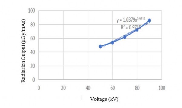
Figure 2: Effect of ESD on Voltage Measured with a Raysafe Multimeter
Figure 2 shows that the effect of radiation output (μGy/mAs) on tube voltage (kV) can be determined from the equation of the number of variables with rank y = 1.0379x 0,9715(μGy/mAs), where x is the tube voltage (kV), and y is the linear regression formula. Then, the score for the Skull AP examination can be obtained by using the linear regression equation. For example, for 10 mAs, 60 kV, and FSD 100 cm, then the ESD value can be determined using the following equation:
So, the ESD value for the Skull AP examination was obtained at 0.0005541 Gy. From the results of the overall calculation, the examination shows that at 40 kV, the smallest ESD value is 0.3737 mGy, while the largest ESD value is 0.7328 mGy at a voltage of 80 kV. It can be explained that the greater the voltage, the greater the ESD value. This result aligns with the research conducted by Duan et al. (2024), which indicates that the dose value (mGy) is influenced by voltage (kV), where the higher the voltage, the higher the dose value received by the patient. According to a similar study conducted by Romodin et al. (2025), explaining that the distribution of ESD values was obtained from the calculation results, for all general radiography examination procedures, the ESD value was obtained from the smallest value of 0.002 mGy and the largest 0.41 mGy. Although the dose value received by patients is relatively low, the difference in dose in one type of examination is quite high. Thus, the indirect ESD measurement method can be used as an alternative to solving problems in the field.
The hereditary effect in this study was obtained from the calculation of ESD values, which were then compared with references (Ibrahim et al., 2020; Elamri et al., 2025). The inheritance effect due to ionizing radiation for generation I was 0.41-0.64% and generation II was 0.53-0.91%. Using equation with the type of Skull AP examination (60 kV), it can be determined that the generation I probability of hereditary effect 1 (PHE1) and generation II probability of hereditary effect 2 (PHE2) can be determined, based on ESD results. The ESD results with the mGy unit were changed to Gy, where the values of PHE1 0.41% = 0.13% and, PHE1 0.64% = 0.08% and, PHE2 0.53% = 0.10%, and PHE2 0.91% = 0.06%. Based on the results of the calculation of the hereditary effect at the voltage of 40 kV, it occurred in generation I with a probability value of 0.04-0.06%, and in generation II it had a probability value of 0.04-0.07%, the lowest. At a voltage of 80 kV in generation I, it has a probability value of 0.11- 0.17%, and in generation 8-0.13%. In accordance with the research II, it has the highest probability value of conducted by Ibrahim et al., (2020), the study did not explain the effect of tension on the occurrence of hereditary effects but only explained the background frequency per 106 births of obtaining hereditary effects. Based on the calculation of hereditary effects for generation I has the highest probability compared to generation II; this is highly dependent on the ability of cells after exposure to radiation (Calabrese et al., 2025). Cells, in general, can repair naturally (Keenahan et al., 2025). The longer the time after exposure to radiation, the more cells have the opportunity to repair the effects of radiation (Jafarian & Hoeschen, 2025). So that the probability of the risk of hereditary effects in the second generation will be smaller (Goudarzi et al., 2024).
Limitation
The limitations of this study include several factors that could affect the generalizability and accuracy of the findings. First, the research was conducted at a single institution, the Bali Academy of Radiodiagnostic and Radiotherapy Engineering, which may not represent the broader population or equipment variations. The study also used only a specific model of X-ray equipment, which may not be consistent with other diagnostic systems in different settings. Additionally, the study focused on a limited range of tube voltages (40-80 kV), which may not fully encompass all possible clinical scenarios. The estimation of hereditary effects relies on the calculation of Entrance Surface Dose (ESD), which may not account for individual patient variations such as age, body composition, or pre-existing health conditions. Finally, the assessment of hereditary effects over generations, while valuable, involves assumptions about cell repair and genetic mutation that may require further empirical validation.
Conclusion
In the examination of patients with conventional radiography, X-ray aircraft output measurements were used, where the ESD value obtained varied from 0.374 mGy to 0.733 mGy. Meanwhile, the voltage of 40 kV (lowest voltage) has produced a probability of hereditary effect of generation I of 0.05-0.09%, generation II of 0.04-0.07%, and a voltage of 80 kV (highest voltage) of the probability of hereditary effect of generation I of 0.11-0.17% and generation II of 0.08-0.13%. The probability of hereditary effects of generation I is greater than that of generation II, because cells in general can repair naturally.
Instruction for AI Assistance Declaration
The author hereby declares that, during the preparation of this manuscript, generative AI tools such as ChatGPT, Microsoft Copilot, and Google Gemini were utilized to assist with language enhancement and grammar correction. Following the use of these tools, the author thoroughly reviewed and revised the content and takes full responsibility for the final version of the manuscript, ensuring its accuracy and adherence to the required academic standards.
Conflict of Interest
The authors declare that there are no conflicts of interest related to this study.
Acknowledgement
Gratitude was expressed to the Physics Study Program, Faculty of Mathematics and Natural Sciences of Udayana University and the Academy Technology of Radiodiagnostic and Radiotherapy (ATRO), Bali, Indonesia for the opportunity to use the Laboratory, Raysafe Multimeter and X-ray aircraft for the smooth and successful of this research.
References
Alomairy, N., Hadi, D., Al-Zaid, A., Fasikh, R., Arif, R., Al-Hazmi, R., ... & Shubayr, N. (2023). Evaluation of the Entrance Surface Doses (ESD) for common diagnostic X-ray examinations. Journal of Radiation Research and Applied Sciences, 16(4), 100754. https://doi.org/10.1016/j.jrras.2023.100754
Andriati, S., Irka, F. H., Firmawati, N., Wahyuni, A., & Fardela, R. (2024). Determination of Radiation Dose Rate in Radiology Installations Using Raysafe X2 Surveymeter. Jurnal Pendidikan Fisika dan Teknologi, 10(2), 458-470. https://doi.org/10.29303/jpft.v10i2.7841
Calabrese, E. J., & Selby, P. B. (2025). More fraudulent history of cancer risk assessment: The US National Academy of Sciences Biological Effects of Atomic Radiation (BEAR) I Genetics Panel used falsified data greatly exaggerating hereditary/cancer risks. Chemico-Biological Interactions, 419, 111640. https://doi.org/10.1016/j.cbi.2025.111640
Chang D S., Lasley F D., Das I J., Mendonca M S., Dynlacht J R., 2021, Stochastic, Deterministic, and Heritable Effects (and Some Radiation Protection Basics), Basic Radiotherapy Physics and Biology, Pages 337–348 https://doi.org/10.1007/978-3-030-61899-5_34
Choudhuri, S., Kaur, T., Jain, S., Sharma, C., & Asthana, S. (2021). A review on genotoxicity in connection to infertility and cancer. Chemico-Biological Interactions, 345, 109531. https://doi.org/10.1016/j.cbi.2021.109531
da Silveira, G. S. B., Neves, L. P., Alves, A. D. S. B. Z., de Souza Santos, W., & Perini, A. P. (2026). Organ and skin dose assessment in neonatal chest radiography using Monte Carlo simulation. Radiation Physics and Chemistry, 113621. https://doi.org/10.1016/j.radphyschem.2026.113621
Duan, Y., Li, X., Lu, P., Yang, C., Wang, K., Zhang, D., ... & Li, B. (2024). High-voltage ESD protection devices with high robustness of 13 kV and strong radiation tolerance up to 200 krad (Si). IEEE Transactions on Electron Devices, 71(6), 3518-3524. https://doi.org/10.1109/TED.2024.3389932
Elamri, N., Tahiri, M., El Baydaoui, R., & Mkimel, M. (2025). Evaluation of radiation dose and cancer risk for paediatric digital radiography in a Moroccan hospital. Radiation Physics and Chemistry, 227, 112352. https://doi.org/10.1016/j.radphyschem.2024.112352
Goudarzi, Y., Monirvaghefi, K., Aghaei, S., Amiri, S. S., Rezaei, M., Dehghanitafti, A., ... & Rajabivahid, M. (2024). Effect of genetic profiling on surgical decisions at hereditary colorectal cancer syndromes. Heliyon, 10(15). https://doi.org/10.1016/j.heliyon.2024.e34375
Ibrahim I. K., Hassan F. F., Abdulkareem N. K. (2020). Effective dose calculation for patients undergoing X-ray examinations in Erbil Hospitals. International Electronic Journal of Medicine, 9(3), 121-123. https://doi.org/10.34172iejm.2020.22
Imaoka, T., Nishimura, M., Daino, K., & Kakinuma, S. (2023). Modifiers of radiation effects on breast cancer incidence revealed by a reanalysis of archival data of rat experiments. Journal of Radiation Research, 64(2), 273-283. https://doi.org/10.1093/jrr/rrac090
Jafarian F and Hoeschen S C., 2025, Low-Dose radiation risk in medicine: a look at risk models, challenges, and future prospects. Zeitschrift für Medizinische Physik, 35(4), 393-400, https://doi.org/10.1016/j.zemedi.2025.07.002
Kanzaki, Y., Kuramoto, T., Takarabe, S., Shibayama, Y., Yoshikawa, H., & Kato, T. (2023). Effect of high-and low-energy entrance surface dose allocation ratio for two-shot dual-energy subtraction imaging on low-contrast resolution. Radiography, 29(1), 240-246. https://doi.org/10.1016/j.radi.2022.11.007
Keenahan, L., Ahsan, M. D., Mecklai, A., & Frey, M. K. (2025). Navigating dual risks: Ovarian cancer prevention and cardiovascular health in patients with hereditary cancer syndromes. Gynecologic Oncology Reports, 61, 101940. https://doi.org/10.1016/j.gore.2025.101940
Liu, Z., Ali, M., Sun, Q., Zhang, Q., Wei, C., Wang, Y., ... & Li, X. (2024). Current status and future trends of real-time imaging in gastric cancer surgery: A literature review. Heliyon, 10(16). https://doi.org/10.1016/j.heliyon.2024.e36143
Mothersill, C., Seymour, C., Cocchetto, A., & Williams, D. (2024). Factors influencing effects of low- dose Radiation exposure. Health Physics, 126(5), 296-308. https://doi.org/10.1097/HP.0000000000001816
Rajeev, R., Mohanty, P., Datta, S. S., & Moitra, P. (2025). Recent advances in point-of-care testing devices for transfusion medicine. TrAC Trends in Analytical Chemistry, 118490. https://doi.org/10.1016/j.trac.2025.118490
Rayan, A. M., Adam, A., Al-Arabi, G., & Ahmed, M. R. (2025). The applications of X-ray technology in medical imaging: advances, challenges, and future perspectives (A review). Journal of Sustainable Food, Water, Energy and Environment, 1(2), 39-61. https://doi.org/10.21608/jsfw.2025.409882.1003
Reis, C. S. D., Gulizia, M., Champendal, M., De Labouchere, S., Sun, Z., & Silva, C. (2023). Plain radiography has a role to play in current clinical practice in Western Switzerland. Journal of Medical Imaging and Radiation Sciences, 54(4), 670-678. https://doi.org/10.1016/j.jmir.2023.08.007
Romodin, L. A., Umnikov, A. S., & Samoilov, A. S. (2025). Biological Reactions under the Combined Action of Ionizing Radiation with Other Factors. Мedical Radiology and Radiation Safety, 70(3), 22-33. https://doi.org/10.33266/1024-6177-2025-70-3-22-33
Stewart, F. A., Akleyev, A. V., Hauer-Jensen, M., Hendry, J. H., Kleiman, N. J., Macvittie, T. J., ... & Wallace, W. H. (2012). ICRP publication 118: ICRP statement on tissue reactions and early and late effects of radiation in normal tissues and organs—threshold doses for tissue reactions in a radiation protection context. Annals of the ICRP, 41(1-2), 1-322. https://doi.org/10.1016/j.icrp.2012.02.001
Suliman, I. I. (2020). Estimates of patient radiation doses in digital radiography using DICOM information at a large teaching hospital in Oman. Journal of Digital Imaging, 33(1), 64-70. https://doi.org/10.1007/s10278-019-00199-y
Tandon, R., & Srivastava, N. (2025). Unravelling exosome paradigm: Therapeutic, diagnostic and theranostics application and regulatory consideration. Life Sciences, 366, 123472. https://doi.org/10.1016/j.lfs.2025.123472
Utami, L. R. W., & Alfiani, N. (2024). Elbow joint radiography examination procedure in post open reduction internal fixation cases in the radiology installation of Kendal District Hospital. Journal of Applied Health Management and Technology, 6(2), 100-105. https://doi.org/10.31983/jahmt.v6i2.11107
Tube Voltage (kV)
Radiation Dosage (µGy)
Radiase Output (µGy/mAs)
Settings
Measured
50
51.10
482.63
48.263
60
60.88
538.93
53.893
70
71.52
618.56
61.856
80
81.81
721.80
72.180
90
91.46
857.90
85.790